Winter, R. et als.
Journal of Plastic, Reconstructive & Aesthetic Surgery, 2019-07-01, Volumen 72, Número 7, Páginas 1084-1090
The segmental paraspinous and intercostal blood vessels form the blood supply and represent the pivot point for the reverse latissimus dorsi flap.
Aim of this study was to confirm the exact location of the blood supply and the most caudal pivot point to assess the suitability of the reverse latissimus dorsi flap for pedicled reconstructions of the trunk as well as sacral area.
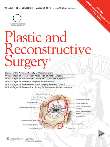 The purpose of this article is to provide a comprehensive review based on images and discussion of the current understanding of the arterial supply of the face to facilitate safe minimally invasive antiaging procedures.
 Burns of anterior shoulder joint surface and neighboring areas produce shoulder edge adduction contracture and scar deformity, slowing down the development of upper limbs in pediatric patients. Therefore, surgical reconstruction is indicated as early as the contracture is formed. Currently used surgical techniques, based on counter transposition of the local triangular flaps and skin transplants, do not solve the problem because of incomplete release of the contractures. Repeated operations are often performed. The scar deformity also remains.
 En este artículo se presenta un heterogéneo conjunto compuesto por 30 prototipos nasales, seleccionados deliberadamente 10 que presentaban perfil con óptima definición del dorso y piel de grosor intermedio con el fin de investigar sistemáticamente, en estos últimos, al segmento inicial del borde superior de la rama alar externa y al tramo distal del borde anterior del cartílago triangular. Mediante disecciones rutinarias, se realizó el análisis con material de individuos adultos, de ambos sexos y raza blanca, previamente formolizado.
 The anatomy of the facial artery, its tortuosity, and branch patterns are well documented. To date, a reliable method of identifying the facial artery, based on surface landmarks, has not been described. The purpose of this study is to characterize the relationship of the facial artery with several facial topographic landmarks, and to identify a location where the facial artery could predictably be identified.




 Sitio web publicado el
Los lectores comentan